[Date Prev][Date Next][Thread Prev][Thread Next][Date Index][Thread Index]
Re:[ccp4bb]: SUMMARY digital imaging of crystals
Dear All,
Many thanks to those who replied to this one.
Original question:
I would like to purchase a system to record images of crystals
electronically. If anyone has come up with a relatively cheap method of
doing this, I would be grateful if they could share their experiences. I
guess the cheapest way is to stick a digital camera on your microscope - we
already have the adaptor for a regular SLR camera. However, I would also
like to hear about other, perhaps more sophisticated solutions.
In the light of some of the responses I should have qualified it by saying I
wanted a system that gave me an instant result. I didn't want to record a
whole tray automatically, just the ones with crystals. Neither did I have a
requirement for sophisticated annotation features. I just wanted to be able
to transfer the images easily to a PC.
Anyway here is a summary of responses:
---------------------------
>From Tassos Perrakis:
Olympous is offering a rather sophistictaed solutionfor a digital
camera. You can catch that signal 'live' via the analog output of the
camera at low resolution (around 600x400) and you can also take still in
high resolution 2048x1536. You have to buy the camera (~ 2000 Euro) the
frame graber for the PC (~500 Euro) - the PC obviously - and some
software from Olympous (which IS necessary to combien the live and
still-high-qulaity capabilities) which is another ~ 1500 Euro.
The alternative we chose (again from Olympous) was to buy from the a JVC
camera for ~1800 Euro for live image and use teh frame grabber to save
images. The quality of that is not oustandign - by any means - but good
enough even for publication in smal lsize - i.e. single column Acta D.
Some free-ware framegrabbers (i.e. IrfanView have capabilities for time
lapse photography. Together with a real 'cold-light' source it can be
fun and educational to take pictures of crystals growing.
Another solution is the Pixera cameras which have some cheaper models
which are fine. You can buy these from Olympous as well or directly from
Pixera. Olympous will be slightly more expensive, but then they gurantee
that the whole boogie works.
--------------------------
>From Harry Powell
Much cheaper (in terms of capital expenditure) and higher quality than a
digital camera would be a flat-bed scanner (no need to spend more than 50
- 100 GBP; if you want to scan 35mm slides as well you can buy an adaptor
for many scanners for an extra 30 - 50 GBP) and continue using your SLR.
Of course, you'd still have the running costs of film, and delays in
processing etc...
This caused a little confusion that was cleared up in a subsequent
message......
Sorry if it wasn't clear from my previous messaage. I meant you scan
photographs (of your crystals) which were taken previously using a film
SLR...
------------------------------
>From Bjorn Kauppi:
We bought a Nikon Coolpix950 last year with an adaptor (sold by nikon) to
our nikon microscope and we are very happy with its performance. It
records the pictures on a flashram card which can easily and fast be
transferred to a computer with an USB port. This is much cheaper than
special high-end digital cameras for microscopes but my feeling is that it
is more than enough for our purposes, with the additional advantage that
it can be used as an normal digital camera as well if you want to document
something in the lab. We also use it for PAGE gels etc.
--------------------------------
>From Bernie Santarsiero:
There are several alternatives.
CrystalScore from www.dsitech.com is one option. They have an automated
stage
and can take one complete set of pics from a crystal plate.
Emerald Biostructures also sells a good digital camera for a microscope, and
a
notebook system for recording and annotation the images.
The basic issues are what are you going to do with the images. Do you want
to
save them all, or just one or two from a crystallization run, or
time-elapsed
images.
The easiest thing to do is get a good digital camera for the microscope,
take
the image, and use photoshop, or some other application like it to modify
and
store the image. Good digital images are about 1MB in size, with enough
resolution to zoom in after the image is collected.
If you talking about saving an entire set of images from a crystal plate,
it's
more complicated, since you have to worry about where the drop is, the zoom
level, focussing, etc.
I hope that helps.
-------------------------------------------
>From Tom Ceska:
I have a video camera (#700) attached to my microscope (Leica) which
is attached to a Matrox video card (#800) on a PC. The system works
reasonably well, and I can capture images to put into Powerpoint
presentations, and also for archiving crystallization tray results.
The system is about 4 years old. I think video cameras cost about
the same, but video capture cards have come down in price a lot.
I am told by Roger Williams that the quality of the picture I get
in the monitor is pretty good and much better than the system he
set up at the MRC (Cambridge).
If you happen to be in the London area, Slough is not very far away,
and you would be welcome to come and have a look at my setup to
see if it meets your requirements.
I got my information about video capture from the microscope
representatives when I bought my microscope. They are of course
interested in selling the most expensive high quality system, but
if pressed they will offer cheaper alternatives. This is what I did.
The risk I had was the unknown quality of the captured image when
I bought the hardware. But I think it is pretty good for almost
everything I want to use the images for.
Regards,
Tom.
-----------------------------------------------
>From Tom Stout:
We bought a "Pixera" camera about 3 years ago....primarily because
it was so afforable (~$1200 at the time which was quite good then).
We still use it, but the old adage is definately true: you get what you
pay for. It is slow & the quality is pretty good at low magnification
(on the scope) for "macroscopic" objects, but when you get down to
the level of most typical protein crystals (100 microns or less), it
doesn't do such a fabulous job. Also, it's purely a manual setup -
no options for auto-scanning trays or dropping all of the images into
a database or anything like that. I can forward you a representative
image if you're at all interested......
On the flip side, I know several people who have bought the digital
microscope cameras from Kodak - there the quality is much higher, but
I understand that it is also much more difficult to use - the images
are stored on the camera until you manually download them to a computer.
The Pixera at least works through a card that you plug into your computer
and images are dropped directly to disk.
-----------------------------------
>From Brent Segelke:
What do you consider cheap? Emerald biostructures sells a system that is
fairly sophisticated and it comes with project management and database
software. DSI also sells a digital microscope camera with similar
capabilities and software support. The DSI system also has crystal detection
software but it gives a lot of false positives. Unfortunately, neither of
these systems is what one would consider cheap. You could probably get a
company to develop a robotic microscope camera for you that would cost less
than the Emerald or DSI systems. Your best bet I'm afraid will be to put a
digital camera on your existing microscope and if you can budget it, have a
x-y stage put on the microscope.
Hope this information is useful. I'd be interested to hear the responses you
get as we are also looking in to digital imaging for crystallization trials.
and....
I guess DSI (http://www.dsitech.com/cscore.htm) would like to see their
product mentioned as well...
There are several groups and must be several companies developing new
devices for automated crystal imaging. I think George DeTitta was fairly far
ahead with a high throughput system for imaging in microbatch a year ago.
----------------------------------------
>From Phil Jeffrey:
Birdwatchers have been doing something analogous for a while - taking
digital pictures from the optics of their (rather high quality) telescopes
("digiscoping"). With digiscoping, often the simple expedient of putting
the digital camera up to the eyepiece and taking the picture will work.
Some tinkering with focus is sometimes necessary. The digital camera's
picture review facility makes life easier.
See: http://www.surfbirds.com/Features/digiscoping.html
as an example. The pictures are surprisingly high quality.
I am guessing that the same approach will work with microscopes as with
telescopes since the optical designs are closely related.
If I might respecfully disagree with Harry Powell, flat bed scanners are
often extremely poor negative/slide scanners. They are especially
atrocious for slides. Much better to get a slide/negative scanner (HP,
Canon, Nikon, Minolta, Poloroid all make respectible models), e.g. the HP
Photosmart S20 gets good reviews. There's a fair amount of www info out
there on the "digital darkroom" if you want to go that route.
and Harry responded......
No need to be respectful about it - I haven't tried the slide/negative
adaptors so can't make any comment about their quality! However, I note
that the US price of the Photosmart S20 is $499 (last updated on HP's
website
http://www.pandi.hp.com/pandi-db/prodinfo.main?product=photos20scanner )
is rather higher than the cost of the slide adaptors I suggested.
You pays your money and you takes your choice...
----------------------------------
>From Quyen Hoang:
Hi,
What we did is similar to what you have, but instead of taking the images
with an "off the shelf" digital camera, we purchased a ccd chip, a
focusing lense and an electronic board. After assembling the components,
we mounted it on a C-mount. We connected the output terminals to a
computer and to a small TV. The TV is used for oberserving the crystals
and the computer is used for capturing and storage. You can use the
computer for observing as well and not need the TV of course.
We also connected a printer to the TV so that a low quality hard copy can
be printed without going through the computer.
Regards,
Quyen
---------------------------------------
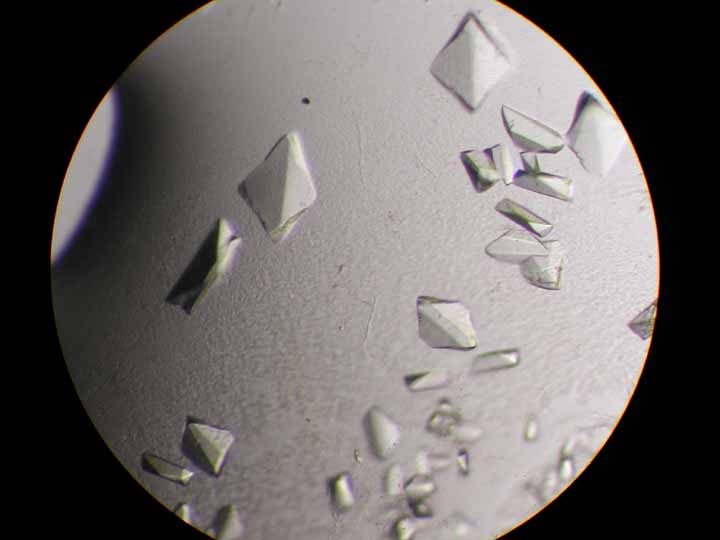
>From Phoebe Rice:
Our students found that you can take any digital camara (ie one meant for
photographing scenery on vacation), hold it just so over the microscope
eyepiece, and shoot quite nice pictures. If you make a little cardboard
adaptor tube that fits over the eyepiece, its even easier. The preview
thingy on the back of the camara is crucial.
The attached pic was taken with my Canon PowerShot A5.
Phoebe
-------------------------------------
>From Lisa Edberg:
I am very happy with our Olympus AX70 Digital microscopy system.
http://www.olympusamerica.com/product.asp?c=21&p=18&s=11&product=47
It has the Olympix 2000 digital camera on it, and DIC optics.
I admit, maybe it was a leetle bit pricey....
I would suggest also getting the lowest power objective available -
sometimes I grow crystals that are too big to photograph!
For crystals grown under oil, you might wish to purchase an inverted
scope.
Lisa
-----------------------------------
>From Ed Berry:
If you're tending toward the high end, I suggest looking into a robotic
microscope stage and crystal tray manipulator so you can give it a
stack of 12 trays and have it take a picture of each well at 0, 1, 6,
12, 24 hr, and daily thereafter; without the necessity of some human coming
into the cold room and breathing moist air all over the lenses. Then
if you solve the structure from the coffin-shaped crystal in well C5
of tray 7, you can go back and make a time-lapse movie of the growth of
that crystal to show in you powerpoint presentation.
And get Emerald or Hampton to mass-produce the system and sell it for
under $10k so we can all get one.
On the low end you can get adapters to put an inexpensive ccd video camera
on the same port used by the film camera, and something like Connectix
"Quick clip" device to grab video or still images from the video stream.
Resolution is lousy, but if you zoom in till the crystal fills the view
its not that bad. Pixera has a digital video system with the same
functionality but refresh rate is much slower than video making it
difficult to focus (at least on slow PC's).
Ed
---------------------------------
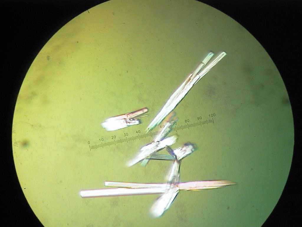
>From Holly Heaslet:
I use a Nikon system. An adapter arm fits between the lens and binoculars.
You
can then place a threaded mount on top of the adapter and screw on your
digital
camera. I use the Nikon Coolpix 990 which runs about $1000. But you can
use
any digital with a threaded lens mount.
I bought my camera from B&H Video http://www01.bhphotovideo.com
For the adapter you can try NikonUSA http://www.nikonusa.com
or Micro Video Instruments http://www.mvi-inc.com
I've attached a baby picture of my crystals that I took with the system.
(See attached file: holly.jpg)
----------------------------------
>From Richard Gillilan:
> There are several groups and must be several companies developing new
> devices for automated crystal imaging. I think George DeTitta was
> fairly far
> ahead with a high throughput system for imaging in microbatch a year
> ago.
>
Incidentally, George DeTitta will be speaking on this topic at the
MacCHESS user workshop June 13. There is a pointer to the
registration page on
www.chess.cornell.edu/Meetings/UserMeeting2001/2001Agenda.htm
------------------------------------
>From Peter Moody:
Our cheap trick is to use the little ccd camera that SG gave away with
indies a few years ago.
------------------------------
Once again, thanks everyone for your contributions,
Dave
-------------------------------
Dr. David M. Lawson
Biological Chemistry Dept.,
John Innes Centre,
Norwich,
NR4 7UH, UK.
Tel: +44-(0)1603-450725
Fax: +44-(0)1603-450018
Email: david.lawson@bbsrc.ac.uk
Web: http://www.jic.bbsrc.ac.uk/staff/david-lawson/index.htm
holly.jpg
phoebe.jpg